
Your doctor has recommended for you to undergo Renal Embolisation. This leaflet will explain what this operation involves, the intended outcomes and the possible risks.
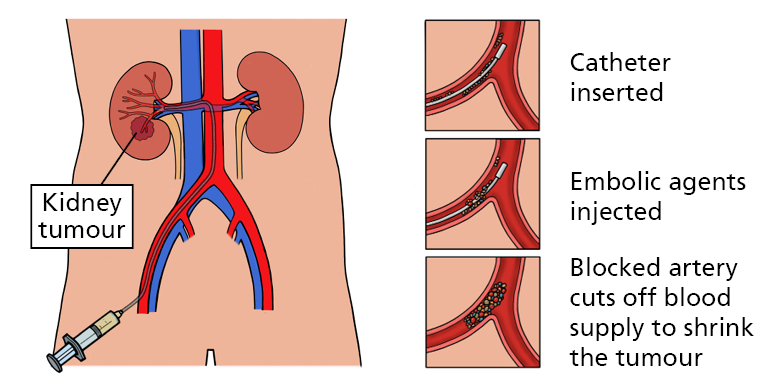
A renal angiogram is done first to look at the blood vessels in your kidneys. This is a special X-ray examination designed to show the vessels to and in your kidneys. A dye (contrast agent) is injected directly into the artery through a fine tube (sheath/catheter) to make the vessels visible on X-ray.
Renal embolisation is when the blood supply to part or whole of your kidneys is purposely stopped. The procedure is done for various reasons such as before surgery to reduce the risk of bleeding, to block blood supply to a kidney tumour, to stop an active bleeding from a blood vessel after trauma, and to support blocking an aneurysmal (ballooned) vessel from rupturing.
Once the interventional radiologist has located your renal blood vessels based on the angiogram, small metal coils or particles or glue are inserted down the catheter and left in the artery. Over time blood clots will form around these foreign material, blocking the blood supply to the part or whole of your kidney.
How do I prepare?
To promote patient safety during your operation, the following patient preparation are required:
- You can eat and drink as normal.
- Most blood thinning medications need to be omitted, please discuss with us if you are taking any.
- If you have bleeding disorders or your blood clotting function is below threshold, you may require a blood transfusion; please inform us if you have concerns.
How is a renal embolisation performed?
Consent
This operation usually takes about 1-1.5 hours, though every patient is different and operations may take longer. The operating interventional radiologist is going to explain this to you in more detail, along with the risks and intended benefits before you provide your permission to go through the operation via a signed consent form.
Anaesthetic
The procedure is performed under local anaesthetic, sometimes supplemented by sedation. Local anaesthetic is used to numb the skin in the groin or the wrist. You may feel a sharp scratch as the anaesthetic goes in but it will feel numb afterwards.
Operation
Once the groin or the wrist is cleaned with an antiseptic and numbed with a local anaesthetic, a needle followed by a wire and sheath (fine plastic tube) are inserted into the blood vessel and passed along your blood vessels to reach the vessels supplying your kidney.
Contrast dye is injected into the blood vessels and X-ray pictures are taken to allow the interventional radiologist to see the area that needs treating. Sometimes this may cause a warm feeling or the feeling that you have passed urine, this is normal. Once the vessels are identified, they are then blocked by inserting small particles or glue or metal coils which act as plugs. Sometimes several arteries need to be blocked, this depends on the blood supply to the area being treated.
Staff involved
Interventional Radiology (IR) procedures are performed by a multidisciplinary team. The team includes the following key Staff:
- Interventional Radiologist (Consultant/Fellow): A doctor specially trained in minimally invasive, image-guided techniques who performs the procedure.
- Radiology Nurses: Highly trained nurses who care for the patient before, during, and after the procedure. They assist with sedation, monitor vital signs (heart rate, breathing, oxygen levels), and may “scrub in” to assist the radiologist directly.
- Radiographers: Experts in imaging equipment who operate the machines to provide real-time guidance for the doctor.
- Senior Clinical Support Workers (SCSWs): Assist with the setup of the room, patient positioning, and maintenance of sterile conditions.
What are the risks
Renal embolisation is a safe operation and like any medical interventions, there are potentially some risks involved.
- Renal infarction: Although all precaution is taken to treat only the area diseased, sometimes surrounding normal kidney tissue (10-25%) can be effected leading to reduced kidney function.
- Post embolisation syndrome usually occurs after the procedure and presents as flu-like symptoms. These will usually resolve in one to two weeks.
- Damage/trauma to the renal artery or on route may occur during the operation and can often be resolved with additional procedures. In rare cases, surgical intervention may be required to restore blood flow especially to the limb or to the kidney. In rare cases, complications can lead to a loss of whole kidney if plan was to treat only part of the kidney at start.
- Patients may experience flank pain during and after the operation; this is normal and can be managed with pain medication but sometimes one may need strong opiods like morphine.
- Rarely after the operation you may experience blood in your urine which should resolve within a week.
- Bruising/Haematoma – Bruising around the site where the needle is inserted into the artery is common and may cause tenderness for a few days. Sometimes, bleeding from the puncture site can lead to a collection of blood under the skin, known as haematoma, which typically resolves on its own.
- A pulsating lump may form in the groin where the tube was inserted, called a pseudoaneurysm or false aneurysm. Treatment may involve a simple injection to block the connection to the artery or, occasionally, minor surgery.
- There is a risk of the dead kidney tissue becoming infected, which can be treated with antibiotics.
- The dye used in this operation can reduce kidney function. Your kidney function will be assessed before the procedure to determine if any additional precautions are necessary.
- Failure to do the operation: As with any medical procedure, there’s a possibility that the intervention may not be successful.
- Allergic reaction to contrast dye (mild symptoms like nausea, itchiness and rashes occur in 3%; moderate to severe symptoms such as severe vomiting, bronchospasm occur in less than 1%; risk of death is rare, estimated at 1:170,000)
- X-rays will be used to make images of your body and guide your doctor during the procedure. X-rays are a type of radiation. We are all exposed to low levels of natural radiation as part of our everyday lives and medical x-rays give an extra dose of radiation.
- Radiation can increase your chances of developing cancer many years or decades after the exposure. The chances of this happening to you as a result of this procedure are considered to be very low. It is important to remember that your doctor thinks that the benefits of this procedure outweigh any risks from the radiation. We will make sure that the amount of radiation used in your procedure is as low as possible.
What to expect from this operation?
Admission
You will be admitted the night before your operation under the care of a renal doctor. At times, you will already have been admitted in a ward and your doctor will arrange:
- Specific and up-to-date blood tests are required to make sure it’s safe to carry out the operation.
- A cannula will be inserted in your arm, and you will need a fluid drip overnight or antibiotics before your operation.
Recovery
After the operation, you will be transferred to our recovery area, where you will be closely monitored:
- Your vital signs and the punctured site will be monitored closely.
- You will be placed on bed rest for several hours to allow you to recover from the operation and reduce your bleeding risk from the punctured wound.
Aftercare
Once stable, you will then be transferred back to the ward to recuperate. You may expect the following:
- You would be asked to drink plenty of water and finish a bag of IV drip to lessen the risk of a kidney injury.
- Your blood tests will also be checked to allow the ward doctor to ascertain when it’s safe for you to be discharged from the hospital.
- If your procedure was to facilitate further surgery on the kidney, then you will progress to that procedure.
Care at home
All being well, you will normally be discharged after 1-2 days.
- Exercise: limit your activities, get plenty of rest and gradually introduce light exercise after a week unless advised otherwise.
- Work: After a week you can return to work.
- Driving/travel: no definite contra-indication for travelling; however, you are advised not to drive for at least 72 hours after your procedure.
- Medicines: medications for pain, sickness and fatigue will be included, when you’re discharged; if you were asked to stop taking blood thinners, we will also include instructions when to resume those.
- Follow-up: attend all follow-up appointments with your healthcare provider to monitor your recovery and assess the effectiveness of the procedure.
- Be aware of signs of complications such as persistent pain, fever, chills, or changes in urination, and contact your G.P Surgery if you experience any of these symptoms.
Contact Information
We hope some of your questions have been answered by this leaflet. If there are any questions you would like to ask before you come for your operation, please get in touch.
Via telephone:
Interventional Radiology SJUH: 0113 326 6841
(Monday – Friday from 9am until 5pm, except Bank Holidays)
Ward 46 SJUH: 0113 326 9146 (Out-of-hours)
Renal Nurse Specialists: 0113 326 7692
Your GP or NHS direct on 111 (routine) | 999 (emergency)
Or via email: [email protected]