
If there are leaks of fluid around the tube, OR pain on feeding, OR new bleeding, OR the gastrostomy tube falls out, stop feed immediately and telephone for urgent advice.
CONTACT WARD J91 AT SJUH on 0113 206 9191 or, 0113 206 8291
This information has been designed to help you understand more about gastrostomy tubes, why they are used, how they are inserted and the care needed afterwards.
It is not meant to replace informed discussion between you and your doctor but can act as a starting point for such discussions.
What is a gastrostomy?
A gastrostomy is a feeding tube placed through your abdomen into your stomach. This tube can be inserted by a number of techniques: using x-ray guidance (RIG), with the help of endoscopy or by surgery.
Radiologically inserted gastrostomies (RIGs) are usually reserved for patients who are unable to have an endoscopic procedure. Surgery is reserved for patients who cannot have the tube inserted by endoscopy or x-ray guidance.
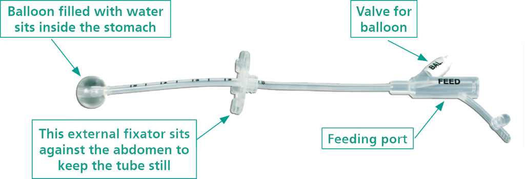
RIGs inserted at Leeds Teaching Hospitals have a balloon at the tip that is blown up by being filled with water. This holds the tube in your stomach.
Why do I need one and what are the alternatives?
If you have difficulty eating and drinking, it may not be possible for you to have all the nutrients and calories you need. The gastrostomy tube allows you to have some, or all the nutrition and fluid you need without having to swallow it. Liquid feed, water and medication can be given directly into your stomach.
If you wish to look into alternatives to a RIG, please contact your doctor for advice.
Where will the procedure take place?
On the day of your procedure, you will be admitted onto a ward, an Admissions Lounge or an Assessment Area. You will be seen by a nurse who will check what medications you are taking, if you have any allergies, and will discuss any relevant past medical history.
The procedure is undertaken in Radiology Theatres. This is like an operating theatre into which specialised x-ray equipment has been installed.
Staff involved:
- Interventional Radiologist: A doctor who is specially trained in this procedure.
- Radiology Nurses: Specialist nurses who will care for you before, during, and after the procedure.
- Radiographers: specialist staff who work with x-ray equipment.
- Senior Clinical Support Workers: Assist with the setup of the room and the procedure.
How is the RIG placed?
Recovery
After the RIG is inserted, you will be taken to the ward where the nurses will continue to monitor you. The tube in your nose will be removed. You will need to have nothing to eat or drink for eight hours after the procedure.
The area around the RIG may feel quite bruised for a few days and you will need to stay in hospital until the Doctors, Nurses and Dietitians are satisfied the RIG is working correctly. You should be prepared to stay in hospital for at least 24 hours after the RIG has been inserted.
If you are to start feeding through the RIG straight away, a feeding regime will need to be established. A supply of feed and equipment will be given to you before you are discharged from the hospital, and an arrangement will be made for this to be delivered to your home regularly.
Exercise
- Limit your activities, get plenty of rest and gradually introduce light exercise after 48 hours.
Work
- If your job is sedentary and involves minimal physical strain, you can likely return after 24 hours, following your doctor’s guidance. If your job involves heavy lifting, climbing stairs, or strenuous activity, wait at least 48 hours and slowly increase activity as you feel ready.
Driving
- You are advised to avoid driving for 24 hours, especially if you were given sedation.
Medicines
- If required when at home, it is best to continue your usual pain relief. If you take a blood thinner medication, the doctor will give advice about when you
Are there any risks?
As with any procedure, there can be complications. Occasionally, it is not possible to place the tube into your stomach. This means that your doctor may discuss a different method of placement.
- Damage to structures surrounding the stomach such as the bowel and the liver is a very rare possibility and an operation may be necessary to address any problems. As x-rays are used to guide the insertion, this complication is often avoided.
- Extremely rarely, a blood vessel can be punctured when passing the needle into the stomach; which can result in bleeding. This may stop by itself, or you may require another procedure to block the bleeding artery or an operation to stop the bleeding. You may also need a blood transfusion.
- An allergic reaction to the X-ray dye (contrast) is rare. Mild symptoms like feeling sick, itchiness and rashes occur in 3% of cases; moderate to severe symptoms such as severe vomiting and breathing problems occur in less than 1% of cases; the risk of death is very very rare and occurs in 0.0006% of cases.
- The level of radiation used during this procedure is equal to the natural radiation we receive from the atmosphere over approximately two years. The risk from this radiation dose is very low.
- In some very rare situations, you might experience some hair loss or some reddening of the skin in the area that was X-rayed. If this happens, you should contact your medical team who will be able to give some advice on the simple steps you can take to look after your skin.
Further Risks
Sometimes, there may be a leak around the tube which can make your skin very red and painful. It may also lead to a general abdominal infection (peritonitis). We will attempt to treat this, but it may become necessary to remove the tube to allow healing to occur. An operation may also be needed to sew up the hole in your stomach.
It is important to remember that your doctor thinks that the benefits of this procedure outweigh any risks from the radiation. We will make sure that the amount of radiation used in your procedure is as low as possible.
What do I need to do to prepare for the procedure?
You will be asked not to eat or drink for six hours before the procedure. You may be asked to have special disinfectant washes in the five days prior to the procedure. You may also be asked to drink a special preparation the night before the procedure. This contains an agent (contrast) which will help the doctor avoid damaging structures around your stomach.
- When in hospital, a small cannula (thin tube) will be placed into a vein in your arm through which you will receive antibiotics.
- If you have any allergies or have previously had a reaction to the x-ray dye (contrast), you must tell your doctors and nurses at the first opportunity.
- Patients who are or might be pregnant must inform a member of staff prior to the procedure.
How long will the RIG stay in?
This depends, if and when, you can swallow safely or take enough nutrition and fluid by mouth. Your Dietitian and Speech and Language Therapist will give you more information about this. Generally, the tube is changed every three to four months. This is a simple procedure and can be taken place by the specially trained nurses at home.
Looking after your RIG
You will be given a separate booklet and training about looking after your RIG. The nursing team will help you look after your RIG and insertion site regularly. They will teach you about the care of the tube, including the importance of maintaining the water inside the balloon. This ‘balloon water check’ will be undertaken every one to two weeks to prevent the tube falling out. The nursing staff will do this for you until you feel confident to do this yourself.
Contact information
We hope some of your questions have been answered by this leaflet. If there are any questions you would like to ask before you come for your operation, please get in touch.
Contact information for Interventional Radiology


How to get to our departments
We are in the Lincoln Wing of S J U H, Ground Floor. Check the link below to see how to get to us.
We are also located in the Jubilee Wing of LGI, Ground Floor.. Check the link below to see how to get to us.
https://www.accessable.co.uk/leeds-teaching-hospitals-nhs-trust/leeds-general-infirmary/access-guides/jubilee-wing
Back to top